My work at the Laboratory of Cognitive Neurobiology at BU Medical was my first foray into research, and an amazing experience. As a Junior in high school, I participated in my high school’s “Authentic Science Research”(ASR) Program. This program allowed a select group of seniors to study of scientific field of throughout the school year in the form of a research class.
At the end of our year of personal study, all members of the program were encouraged to go and pursue an internship in their selected area of study. For my experience in ASR, I choose to study clinical neurobiology, and was fortunate enough to have Professor Douglas Rosene accept me in for a summer internship in his laboratory.
The Lab
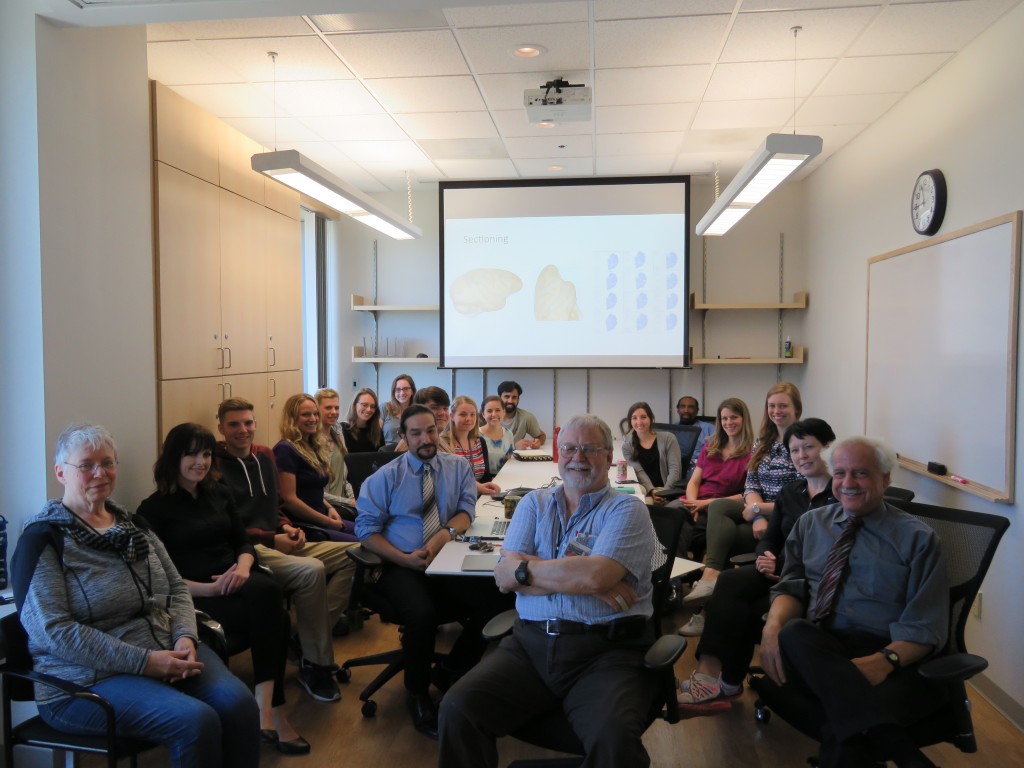
The Laboratory of Cogntive Neurobiology focuses the majority of their research efforts on the neuological bases for learning and memory in non-human primates (NHPs), with a particular interest in aging and age-related disease. The lab uses Rhesus monkeys as a model for human aging and age-related disease, using behavioral data and brain-tissue analysis to uncover neurobiological mechanisms under study. For more information, check out their homepage.
Learning the Ropes
Under Dr. Rosene, I learned the ropes of working with tissues, learning key lab techniques, and better familiarizing myself with NHP brain anatomy. After this preliminary training was complete, I began work on two projects.
Chronic Traumatic Encephalopathy
Dr. Rosene was kind enough to invite me to sit in and contribute my thoughts during several meetings between reseracher Ann McKee and her associates. McKee was in the process of compiling information regarding CTE in NFL players, other athletes, and former war veterans who had been exposed to blast trauma. I aided in her research throughout the summer by processing CTE brain tissue samples - conducting immunohistochemical assays in order to collect data on the expression and localization of disease-related proteins.
In addition to the lab skills I gained through aiding McKee’s work, I found that sitting in on her meetings on experimental design and disease analysis were extremely enlightening. Listening to such bright minds was helpful to me in understanding scientific inquiry at a higher level and better understanding the process of experimental design in the real world.
About a year after participating in this work, the study I worked on under McKee was published and shook the media, leading to a much-needed publicity crisis for NFL and greater public awareness about the disease. To read more about this study, check out this post from The Brink.
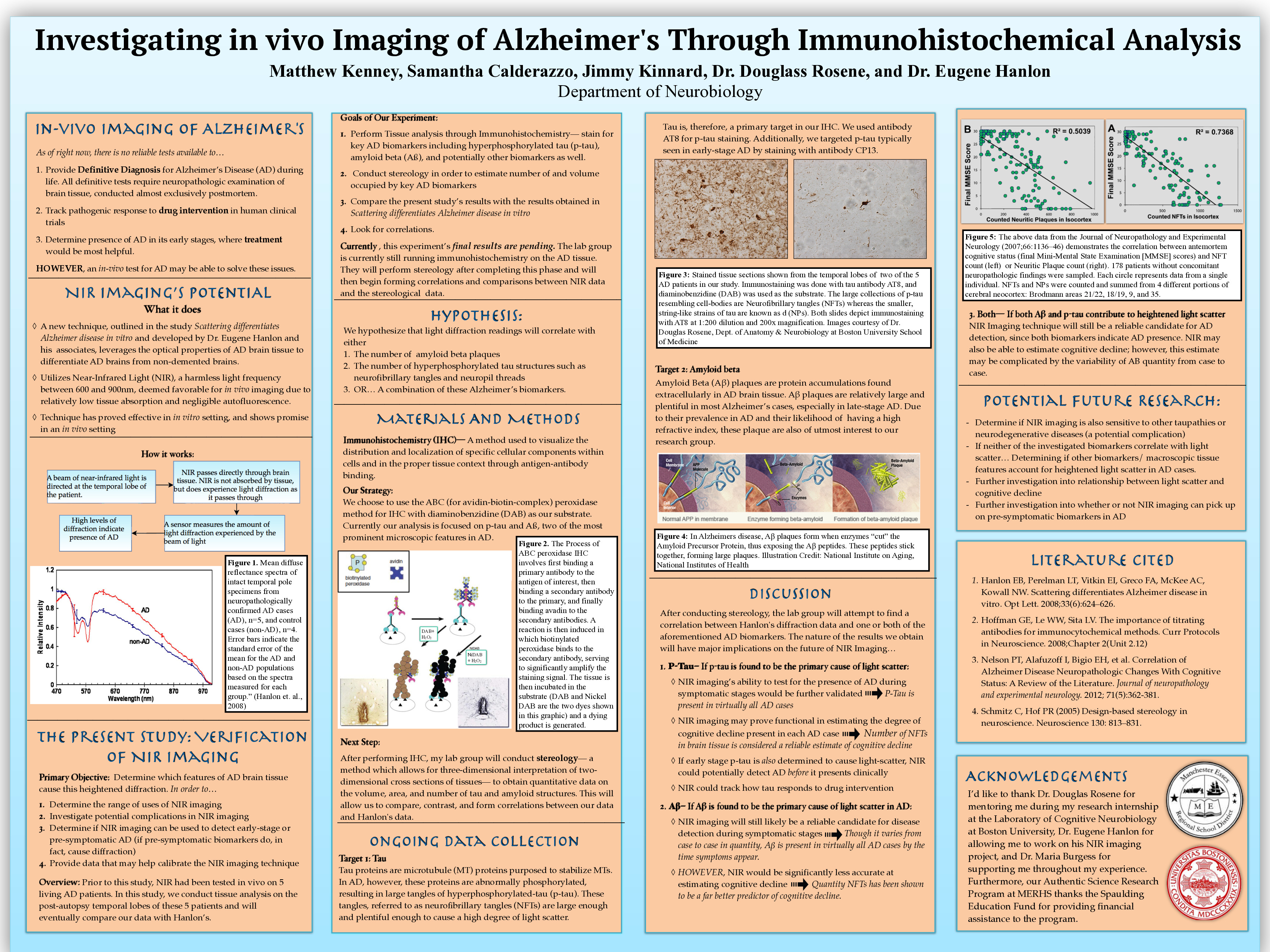
Near-Infrared Light Detection of Alzheimer’s Disease
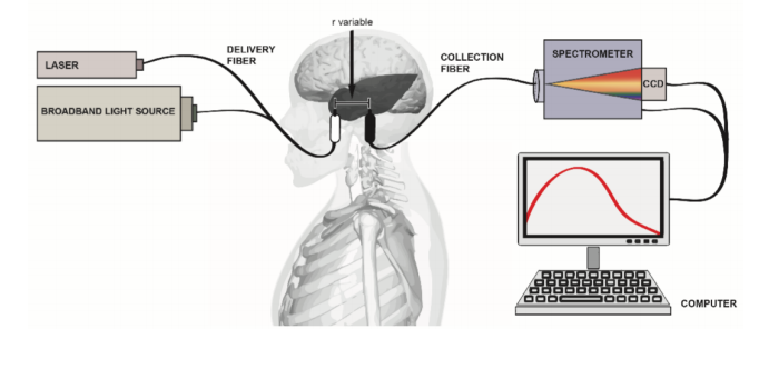
This project consitituted my primary commitment throughout the summer. For this project, I worked on a team with 2 graduates students, under the mentorship of Dr. Douglas Rosene, to validate and calibrate a new Alzheimer’s detection technique. Our work was focused on validating the results of Scientist Dr. Eugene B. Hanlon (and his associates’) new technique which utilizes near-infrared light for in-vitro detection of Alzheimer’s Disease.
The new optical technique created by Hanlon involves passing near-infrared light, a harmless wavelength of light, through brain tissue. Once the light passes through, a sensor at the opposite end of the tissue measures the amount of light diffraction that occured during the pass-through. Alzheimer’s Brain tissue has unique morphological properties (existence of hard plaque buildups, atrophy, etc.) which cause the light-diffraction profile to differ from ordinary brains.
My team’s job was to determine which particular features of the Alzheimer’s tissue were responsible for changes in the light diffraction profile. We conducted immunohistochemical assays and stereology on Alzheimer’s and control brain tissue throughout the summer to zero-in on these features.
End-Goal
If we could determine what features were causing the abnormal light-diffraction profile in Alzheimer’s tissue, we could identify whether or not the abnormal diffraction profile would be consistant across all Alzheimer’s patients. Further, we could instruct Dr. Hanlon to fine-tune his detection mechanism.
The ultimate goal of the experiment was to improve the detection technique to the point where it could be used to detect the presence of Alzheimer’s disease in living people, ideally before the presentation of clinical memory impairments. Such a tool could allow Alzheimer’s patients to take preventative measures prior to developing the full disease symptomology. This project remains ongoing, and has been under active development long after I left the lab at the conclusion of my internship
Reserach Presentation
As a part of my Authentic Science Research program, I presented my research at the JSHS high school STEM compeition, run by the Department of Defense. To get a closer look at my research, click my poster below, or read my paper summarizing the reserach project.